Vascular Lesion 微細血管腫脹,曲張
Vascular lesions include acquired lesions (eg, pyogenic granuloma) and those that arise at or shortly after birth (vascular birthmarks). Vascular birthmarks include vascular tumors (eg, infantile hemangioma) and vascular malformations. Vascular malformations are congenital, life-long, localized defects in vascular morphogenesis and include capillary (eg, nevus flammeus), venous, arteriovenous (eg, cirsoid aneurysm), and lymphatic malformations. Vascular birthmarks usually involve only the skin and subcutaneous tissues but rarely affect the CNS.
Infantile Hemangioma
Infantile hemangiomas are raised, red or purplish, hyperplastic vascular lesions appearing in the 1st year of life. Most spontaneously involute; those obstructing vision, the airway, or other structures require treatment, usually with oral corticosteroids. Surgery is rarely recommended.
Infantile hemangiomas (IH) can be classified by appearance as superficial, deep, cavernous, or by descriptive terms (“strawberry hemangioma”), but all share a common pathophysiology and natural history; therefore, the inclusive term infantile hemangioma is preferred. They are the most common tumor of infancy, affecting 10 to 12% of infants by 1 year.
IH are present at birth in 10 to 20% of those affected and almost always within the 1st several weeks of life; occasionally, deeper lesions may not be apparent until a few months after birth. Size and vascularity increase rapidly, usually peaking at about age 1 year.
Superficial lesions have a bright red appearance; deeper lesions have a bluish color. Lesions can bleed or ulcerate from minor trauma; ulcers may be painful. IH in certain locations can interfere with function; those on the face or oropharynx may interfere with vision or obstruct the airway; those near the urethral meatus or anus may interfere with elimination. A periocular hemangioma in an infant is an emergency as even a few days of disruption of vision can result in permanent visual defects. Lumbosacral hemangiomas may be a sign of neurologic or genitourinary anomalies.
Lesions slowly involute starting at 12 to 18 month, decreasing in size and vascularity. Generally, IH involute 10%/year of age, (eg, 50% by age 5, 60% by age 6), with maximal involution by age 10. Involuted lesions commonly have a yellowish or telangiectatic color and a wrinkled or lax fibrofatty texture; residual changes are almost always proportional to the lesion's maximal size and vascularity.
Diagnosis and Treatment
Diagnosis is clinical; the extent can be evaluated by MRI if lesions appear to encroach vital structures.
Treatment is controversial. Many physicians treat lesions early to prevent subsequent enlargement or to make them less noticeable; others do not treat unless a lesion causes (or risks) functional problems by its location. When treatment is elected, laser treatment or intralesional or systemic corticosteroids are chosen based on the location, extent, and rate of growth of the lesion. For systemic corticosteroid therapy, prednisone 1 to 3 mg/kg po bid or tid is given for ≥ 2 wk. If resolution starts, the prednisone should be decreased slowly; if not, the drug should be stopped.
Topical treatments and wound care are useful for ulcerated lesions and help prevent scarring, bleeding and pain. Compresses, topical mupirocin or metronidazole, barrier dressings (polyurethane film dressing or petrolatum-impregnated gauze), or barrier creams may be used.
Unless complications are life threatening or vital organs are compromised, surgical excision or other destructive procedures should be avoided because they frequently result in more scarring than occurs with spontaneous involution. To help parents accept nonintervention, the physician can review the natural history (photographic examples are helpful), provide serial photography of the lesion to document involution, and provide a sympathetic ear to parents' concerns.
Nevus Flammeus and Port-Wine Stain
Nevus flammeus and port-wine stains are capillary vascular malformations that are present at birth and appear as flat, pink, red, or purplish lesions.
Nevi flammei are flat pink marks that are very common on the nape, glabella, and eyelids. Lesions around the eyes disappear in a few months. Nape lesions may disappear in early childhood, only to recur in middle age.
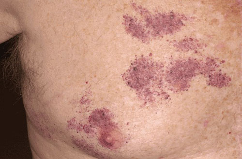
Port-wine stains are flat, reddish to purple lesions appearing anywhere on the body. Lesions become darker and more palpable with time (often becoming quite hyperplastic by late middle age), although the lateral extent does not enlarge beyond the growth of the patient. Port-wine stains of the trigeminal area may be a component of the Sturge-Weber syndrome (in which a similar vascular lesion appears on the underlying meninges and cerebral cortex and is associated with epilepsy).
Diagnosis is clinical. Treatment with vascular lasers produces excellent results in many cases, especially if treated as early in life as possible. The lesion can also be hidden with an opaque cosmetic cream prepared to match the patient's skin color.
Neuvs Araneus
(Spider Nevus; Spider Angioma; Vascular Spider)
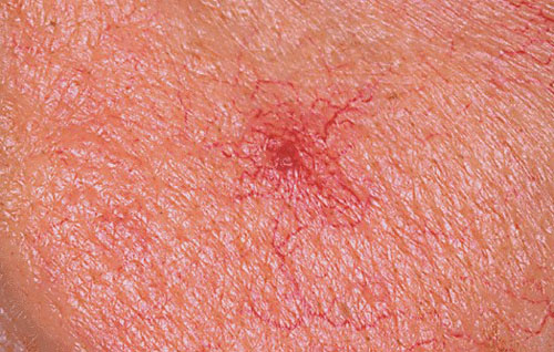
Nevus araneus is a bright red, faintly pulsatile vascular lesion consisting of a central arteriole with slender projections resembling spider legs.
Compression of the central vessel temporarily obliterates the lesion. Lesions are acquired. One lesion or small numbers that are unrelated to internal disease may occur in children or adults. Patients with hepatic cirrhosis develop many spider angiomas that may become quite prominent. Many women develop lesions during pregnancy or while taking oral contraceptives. The lesions are asymptomatic and usually resolve spontaneously about 6 to 9 mo postpartum or after oral contraceptives are stopped. Lesions are not uncommon on the faces of children.
Diagnosis is clinical. Treatment is not usually required. If resolution is not spontaneous or treatment is desired for cosmetic purposes, the central arteriole can be destroyed with fine-needle electrodesiccation; vascular laser treatment may also be performed.
Pyogenic Granuloma
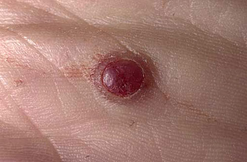
Pyogenic granuloma is a fleshy, moist or crusty, usually scarlet vascular nodule composed of proliferating capillaries in an edematous stroma.
The lesion, composed of vascular tissue, is neither of bacterial origin nor a true granuloma. It develops rapidly, often at the site of recent injury (although injury may not be recalled), and probably represents a vascular and fibrous response to injury. There is no sex or age predilection. The overlying epidermis is thin, and the lesion tends to be friable, bleeds easily, and does not blanch on pressure. The base may be pedunculated and surrounded by a collarette of epidermis. The lesions occasionally resemble and must be differentiated from melanomas or other malignant tumors. During pregnancy, pyogenic granulomas may become large and exuberant (eg, gingival pregnancy tumors, or telangiectatic epulis).
Diagnosis involves biopsy and histologic examination. Treatment consists of removal by excision or curettage and electrodesiccation, but the lesions may recur.
Lymphatic Malformations
(Lymphangioma; Lymphangioma Circumscriptum; Cystic Hygroma; Cavernous Lymphangioma)
Lymphatic vascular malformations are elevated lesions composed of dilated lymphatic vessels.
Lesions are usually yellowish tan but occasionally reddish or purple if small blood vessels are intermingled. Puncture of the lesion yields a colorless or blood-tinged fluid. Diagnosis is made clinically and by MRI. Treatment is usually not needed. If excised, recurrence is common even when there is extensive removal of dermal and subcutaneous tissues.